
The Fallopian Tubes
The only female sex organs whose only biological purpose seems to be ...
reproduction
Dr. Nelson Soucasaux , Brazilian gynecologist
Despite their beauty and delicacy, of all the female sexual organs the
Fallopian tubes [see drawings, below] are the only ones whose only biological
purpose seems to be ... reproduction (at least as far as we know). From
the point of view of interdisciplinary studies, unfortunately it is also
very difficult to find archetypal references to these organs in mythology.
Both from the structural and functional points of view, the Fallopian tubes are
extremely delicate organs and, therefore, highly vulnerable to all kinds
of aggressions, mostly infectious/inflammatory processes that easily may cause
their obstruction and/or impairment of their movement and function, resulting
on infertility or tubal pregnancies.
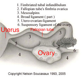
In almost all their length, each Fallopian tube is formed by a thin
cylindrical muscular layer internally lined by a folded mucosa named the
endosalpinx. From the point of their uterine insertion on, the tubes follow
their own way towards the ovaries. Throughout their path, they run along
the broad ligaments, which they are attached to by means of peritoneal folds
named mesosalpinges.
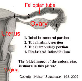
Three portions can be distinguished in the Fallopian tubes: the intramural,
the isthmic and the ampullary.
The intramural portion consists of an extremely narrow canal that crosses
the uterine wall for opening into the uterine (endometrial) cavity.
The isthmic portion starts just at the point where the tubes emerge from
the uterine upper angles; it is less narrow than the intramural and, as
it approaches the ampullary portion, it gradually widens. (There is no sharp
distinction between the isthmic and the ampullary portions.) As to the ampullary
portion, Dr. Frank Netter observes that it is "... tortuous and gradually
widens toward the outer end. It terminates in a fimbriated infundibulum
which resembles a ruffled petunia or sea anemone. On the fimbriae, the fimbria
ovarica is grooved and runs along the lateral border of the mesosalpinx
to the ovary."* Further on: "Contraction of the longitudinal muscle
fibers of the ovarian fimbria brings the infundibulum in close contact with
the surface of the ovary."* This seems to be the main mechanism responsible
for the "embrace" given to the ovary by the tubal infundibulum
at the moment of ovulation, with the purpose of capturing the oocyte [egg]
that is being expelled.
 |
 |
The tubal muscular layer is formed by an intricate arrangement of circular
and longitudinal smooth fibers, facilitating the propagation of the tubal
peristaltic waves towards the uterus. The tubal circular muscle fibers communicate
with the double system of spiral fibers which, symmetrically entwined and
interlaced around the uterine cavity, constitute most of the myometrium
(the uterus's powerful muscular layer). The fact that the uterine contractile
waves originate in the upper angles of this organ, just at the insertion
of the tubes, is due to this communication between the tubal and uterine
circular and spiral smooth muscle fibers.
The endosalpinx is characterized by the formation of an extremely intricate
system of longitudinally arranged folds. Whereas in the intramural portion
these folds are sparse and small, from the isthmic portion on up to the
infundibulum they gradually become more and more numerous, extremely branched
and arborescent. As Netter observes, in the ampullary portion the configuration
of this endosalpinx folding acquires labyrinth-like features.*
The endosalpinx is lined by a single-layered columnar and ciliated epithelium,
basically formed by two kinds of cells: the ciliated and the secretory ones,
which alternate irregularly along the mucosa. These cells are sensitive
to the ovarian hormones and, according to Ham, their heights, relative or
absolute, vary in accordance to the several periods of the menstrual cycle.**
Netter observes that "... the height of tubal epithelium reaches its
peak during the period of ovulation and is lower during menstruation."*
According to this author, the number of secretory cells becomes greater
along the luteal phase of the cycle.*
Just like the peristaltic movements of the tubal muscles, the ciliar
movement of the endosalpinx (which is due to its ciliated cells) is also
directed towards the uterus. When pregnancy occurs, both of them aim to
carry the fertilized egg to the uterine cavity. On the other hand, the secretory
cells are in charge of supplying nourishment to the egg during this transportation,
which usually takes about 5 to 7 days.
As I have observed in my article "Fundamentos para o Estudo das
Influências Neurovegetativas em Ginecologia" ("Basis for
the Study of the Neurovegetative Influences in Gynecology")***, regardless
of the fact that the tubal motility is fundamentally stimulated by the estrogens,
the simultaneous existence of a co-ordinating activity on the part of the
vegetative innervation of the tubes in this process seems to be evident.
During ovulation, even knowing the crucial role played by the pituitary
LH ovulatory peak in helping the process of oocyte capture by the Fallopian
tube and also the increase in the tubal motility caused by the high estrogen
levels of this phase of the cycle, I still believe it is hard to explain
only endocrinally the authentic "embrace" given in the
ovary by the fimbriated tubal infundibulum in order to so perfectly capture
the oocyte. As already mentioned, it seems to be the contraction of the
Fallopian tube's ovarian fimbria that makes the tubal infundibulum
come closer to the ovary and finally surround it with its fimbriae. In this
way, the possibility of a subtle neurovegetative co-ordination of this extremely
intricate process, as well as of the tubal peristaltic waves that aid to
carry the fertilized egg towards the uterus, must also be seriously considered.
* Netter, F.H. - "The Ciba Collection of Medical Illustrations
- Vol 2, Reproductive System" - USA, 1954.
** Ham, A.W. - "Histology" - J.B. Lippincott Company, Philadelphia,
1965.
*** Soucasaux, N. - "Fundamentos para o Estudo das Influências
Neurovegetativas em Ginecologia" ("Basis for the Study of the
Neurovegetative Influences in Gynecology") - in: Jornal Brasileiro
de Medicina, vol 57, nº 4, October 1989.
The text above is an excerpt from my book "Os Órgãos
Sexuais Femininos: Forma, Função, Símbolo e Arquétipo"
("The Female Sexual Organs: Shape, Function, Symbol and Archetype"),
published by Imago Editora, Rio de Janeiro, 1993. For information on the
book, see page http://www.nelsonginecologia.med.br/orgaos.htm
, at my web site www.nelsonginecologia.med.br
.
Copyright Nelson Soucasaux 1993, 2005
_______________________________________________
Nelson Soucasaux is a gynecologist dedicated to clinical, preventive
and psychosomatic gynecology. Graduated in 1974 by Faculdade de Medicina
da Universidade Federal do Rio de Janeiro, Brazil, he is the author of several
articles published in medical journals and of the books "Novas Perspectivas
em Ginecologia" ("New Perspectives in Gynecology") and
"Os Órgãos Sexuais Femininos: Forma, Função,
Símbolo e Arquétipo" ("The Female Sexual Organs:
Shape, Function, Symbol and Archetype"), published by Imago Editora,
Rio de Janeiro, 1990, 1993. He has been working in his private clinic since
1975.


